Q
Which of the following investigation findings is LEAST likely to be found with the following X-ray abnormality?
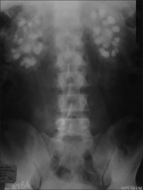
"Photograph shows straight X-ray of abdomen with bilateral extensive nephrocalcinosis."
© Chakrabarti et al.
[Source]
Correct Answer:
Explanation
The X-ray here shows nephrocalcinosis. This is widespread calcium deposition within the renal parenchymal tissue. There are a number of causes which can be broken down into:
Hypercalcaemia and Hypercalciuria:
- Primary Hyperparathyroidism - most common cause (Option E)
- Vitamin D therapy / excessive vitamin D ingestion (Option C)
- Sarcoidosis (Option B)
- Milk-Alkali syndrome
Hypercalciuria without hypercalcaemia:
- Distal renal tubular acidosis - second most common cause
- Medullary sponge kidney
- Inherited tubulopathies
There is some debate about whether nephrocalcinosis is specific to calcium phosphate deposition. Conditions with hyperphosphaturia (+/- hyperphosphatemia) are also therefore implicated
- Tumour lysis syndrome
- Inherited tubulopathies
Calcium phosphate stones form more readily in alkaline urine (Option A).
High oxalate such as in primary hyperoxaluria can be associated with calcium oxalate deposition within kidney parenchymal tissue.
A positive urine nitroprusside-cyanide spot test would indicate cystinuria which although causing a high volume of urinary stones to form would not cause nephrocalcinosis. Additionally cystinuria stones are not high density as seen on the X-ray above.